How do loading dyes differ from DNA stains?
A common point of confusion for students learning DNA gel electrophoresis is understanding the difference between the reagents that aid in visualization. What distinguishes a DNA stain from a loading dye? Let’s talk about the functionality of these two important types of molecules…
What is a gel loading dye?
A gel loading dye is a colored dye that is mixed with a DNA sample prior to running electrophoresis. Loading dyes serve a few purposes:
- They color your sample, making it easier to tell if you were successful in loading your gel.
- They are dense and keep the DNA in your sample from diffusing away in the buffer.
- They migrate through the gel alongside the DNA, helping you track the rate of migration of your sample through the gel.
The image above shows loading dyes migrating through a gel during an electrophoresis run (sped up 300x). This loading dye is actually a cocktail of two different loading dyes that run at different speeds. Loading dye cocktails like this one are more versatile than individual dyes, as they allow you to accurately track both large DNA fragments, which run slowly, and small fragments, which run much faster. Some common loading dyes you may be familiar with include bromophenol blue and xylene cyanol.
Loading dyes help you see your sample when you load it into a gel, but they don’t allow you to see the DNA fragments themselves. DNA is colorless and invisible to the naked eye, so in order to identify the DNA fragments in your sample, you’ll need to use a DNA stain. DNA stains are pigmented or fluorescent molecules that intercalate, or interact directly with DNA, allowing us to see exactly where the DNA is in the gel.
What is a DNA stain?
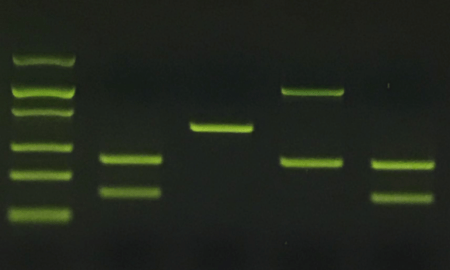
There are many different types of DNA stains. Some are plainly visible in white light, while others require specialized blue light or UV light for visualization. Some are safe for use in classrooms, while others are considered hazardous and require special handling. In the image below, GelGreen®, a fluorescent DNA stain, is used to label DNA bands as they migrate through a gel (sped up 300x). This image was captured under blue light and taken through an orange filter in a blueGel™ electrophoresis system. Another excellent nucleic acid stain is our newer SeeGreen™.
For more information on different DNA stains, see our recent post Choosing a DNA stain for classroom use.