How to visualize biomolecules right in the palm of your hand
Wishing you had something besides paper models or simulations to show how DNA, RNA, and proteins work? With our P51™ Molecular Fluorescence Viewer, you can experiment with those molecules in a simple, visual way. This guide will help you bring this tiny but powerful tool into your classroom.
What is fluorescence?
Fluorescent molecules absorb light energy from specific wavelengths of light and then release some of that energy back out as light of a different color with a longer wavelength. Because these molecules are emitting light rather than simply reflecting it, they appear to glow.
Many molecules found in nature fluoresce, like those found in jellyfish and sea anemones. Scientists have harnessed such molecules to visualize structures and processes that are otherwise undetectable. And with the P51, you can bring that technique to your lab or classroom!
What is the P51 Molecular Fluorescence Viewer?
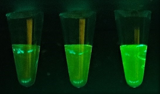
The P51 is a handheld fluorescence viewer designed to enable scientists and students to explore scientific phenomena by direct naked-eye visualization of molecules and their functions. See DNA, RNA, proteins, and more light up to better understand how they work!
Traditionally, when scientists study molecules in the lab using fluorescence, it requires complex and expensive equipment. With the P51, you can use this technology right in the palm of your hand!
How does the P51 viewer work?
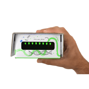
Simply place your samples inside the P51, turn on the blue light, and see your tubes light up! The LEDs in the P51 shine safe blue light to excite fluorescent molecules in your tubes. The yellow or orange acrylic filter in the front filters out the bright blue light so you can see the emitted fluorescence from the excited molecules. The whole system is housed in a sturdy cardboard box, making it both affordable and durable.
As for what kind of molecules you can observe with the P51, we have also developed a whole suite of engaging and innovative curriculum and hands-on lab activities that help students understand how these biological molecules work (see table below).
See the P51 in action:
What can I do with the P51 Molecular Fluorescence Viewer?
We offer a variety of accompanying P51 Molecular Glow Labs compatible with the P51 Fluorescence Viewer that cover a variety of biology topics and techniques, including:
| Learning Lab | Molecule visualized | Techniques | Topics |
|---|---|---|---|
| DNA Glow Lab | DNA (single vs. double-stranded) | Micropipetting Fluorescence detection |
DNA structure Hydrogen bonds Base pairing |
| Enzyme Lab: β-Gal Glow™ | Products of enzyme-catalyzed reactions | Micropipetting Fluorescence detection |
Enzymes Reaction rates Competitive inhibition |
| Free (!) Chlorophyll Lab | Chlorophyll | Paper chromatography | Photosynthesis Environmental science |
| BioBits®: Central Dogma | RNA, proteins | Micropipetting Cell-free technology |
Transcription/translation Central Dogma Protein synthesis Gene expression Synthetic biology |
| BioBits®: Protein Structure and Function | Proteins | Micropipetting Cell-free technology Bioinformatics |
Levels of protein structure Protein structure/function Protein synthesis Protein analysis |
| Introduction to Fluorescence | Fluorescent marker ink | Micropipetting Serial dilutions Standard curves |
Fluorescence Biotechnology Concentrations Scientific notation |
| COVID qPCR Lab | Fluorescent results from qPCR test | Micropipetting PCR/qPCR Gel electrophoresis (optional) |
Infectious disease Molecular diagnostics |
What else do we need to start using the P51 Molecular Fluorescence Viewer?
The additional materials you will need depends on the P51 lab, but generally, you will need:
-
- One P51 Fluorescence Viewer per lab group
- Bundle and save with our classroom set here!
- Power (via one 9V battery or USB-C port)
- 0.2 mL tubes: we recommend these small tubes to dispense and distribute reagents to students because they fit perfectly in the P51
- One 2-20 µl micropipette per lab group, plus micropipette tips
- Fixed volume micropipettes are also sufficient for certain P51 labs, such as the BioBits @home: Central Dogma lab. More about micropipettes in this blog post.
- One P51 Fluorescence Viewer per lab group
Can I use the P51 at home?
Yes! If you want to observe fluorescent molecules at home, our P51 @home: STEM Explorations That GLOW! or BioBits @home: Central Dogma kit has you covered.
Bonus trivia! Why is it called the P51?
In 1952, an X-ray diffraction image of the DNA double helix was made under the direction of Dr. Rosalind Franklin. That important image, known as Photo 51, led scientists to the discovery of DNA’s molecular structure. The P51 fluorescence viewer honors Dr. Franklin’s pioneering work, and provides students and scientists alike with a way to visualize important molecules and biological processes.

Related resources:
- miniPCR bio YouTube: watch how to do all the P51™ labs with our demo videos
- miniPCR bio store: all P51™ products and labs