Choosing a DNA stain for classroom use
Have you ever wondered which DNA stain is best for your gel electrophoresis needs? If so, this blog post is for you!
Because DNA is not visible on its own on an agarose gel, scientists use DNA stains to visualize their samples during or after gel electrophoresis. There are many types of DNA stains, each with different advantages and disadvantages. For classroom use, the most important factors in selecting a DNA stain are safety, ease of use, and cost. Another factor to consider is sensitivity, or how little DNA can be detected by a stain.
Fluorescent stains
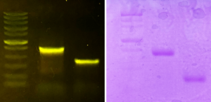
Fluorescent stains are the most commonly used DNA stains in research labs because they are very sensitive, meaning they can detect small amounts of DNA. Fluorescent stains are easy to use and are typically added to the gel when it is being poured. DNA samples are visible as soon as the gel starts to run, and if you use an integrated gel electrophoresis and visualization system (such as the blueGel™ electrophoresis system), your students can even watch their samples separate in real time. However, fluorescent stains can be more expensive and require specialized equipment to excite and visualize the fluorescent dye.
- UV stains: Because UV light sources require additional safety precautions, UV stains are becoming less common in classroom settings. The most common UV-excited DNA stain is ethidium bromide. However, like UV light, ethidium bromide is also hazardous, requiring extra safety precautions and special disposal, and many educational settings prohibit its use.
- Blue light stains: For safety reasons, many scientists and teachers prefer to use DNA stains that are excited by safe blue light. There are numerous commercially available stains of this type, including SYBR® Safe, GelGreen®, and SeeGreen™. Newer blue light stains are much improved over their early predecessors with increased sensitivity and with the ability to resist photobleaching in ways that rival their UV counterparts.
.
Comparison of blue light fluorescent stains
| Advantages | Disadvantages | |
|---|---|---|
| SYBR | Does not distort migration patterns | Photobleaches easily |
| GelGreen® | Highly photostable | Interacts strongly with DNA distorting band appearance |
| SeeGreen™ | Highly sensitive, stable, faithful Compatible with blue and UV light |
Migrates towards cathode, not ideal for long gel runs (>40 minutes) |
Colorimetric stains
There are numerous DNA stains that mark DNA with a visible blue compound (e.g. FlashBlue™, CarolinaBLU®, Fast Blast). Colorimetric stains are more affordable than fluorescent stains and technically do not require a transilluminator (although a white light illuminator is recommended for easy viewing). However, colorimetric DNA stains come with a few major disadvantages. First, colorimetric stains are less sensitive and cannot detect lower concentrations of DNA reliably. This makes them suitable only for some educational uses but not for most real-world lab applications. Second, colorimetric stains involve extra steps and do not allow students to see their results immediately after a gel electrophoresis run. Gels need to be soaked in blue DNA stains and then rinsed, with the staining and destaining process taking at least 15 minutes and up to overnight. The stains are also somewhat messy and can leave blue residue on surfaces.
| Advantages | Disadvantages | |
|---|---|---|
| Fluorescent stains |
|
|
| Colorimetric stains |
|
|
In my experience as a former scientist and classroom teacher, fluorescent DNA stains are worth the increased cost. They are sensitive enough to ensure that students can visualize their DNA samples, even when the concentration is low. Plus, class time is precious and avoiding the soaking and rinsing steps means students get more time to devote to more meaningful educational activities. You can even buy convenient all-in-one tabs that contain agarose, buffer, and fluorescent DNA stain to make casting gels a breeze!
Contributed by miniPCR bio curriculum specialist Allison Nishitani, PhD


